
Conjunctivopalpebral fistula as an unusual complication after ptosis surgery
successful management with fractional CO₂ laser
DOI:
https://doi.org/10.70313/2718.7446.v18.n4.462Keywords:
eyelid ptosis, ptosis surgery, conjunctivopalpebral fistula, CO2 laserAbstract
Objective: To describe the therapeutic management of a patient with a persistent conjunctivo-palpebral fistula following ptosis surgery.
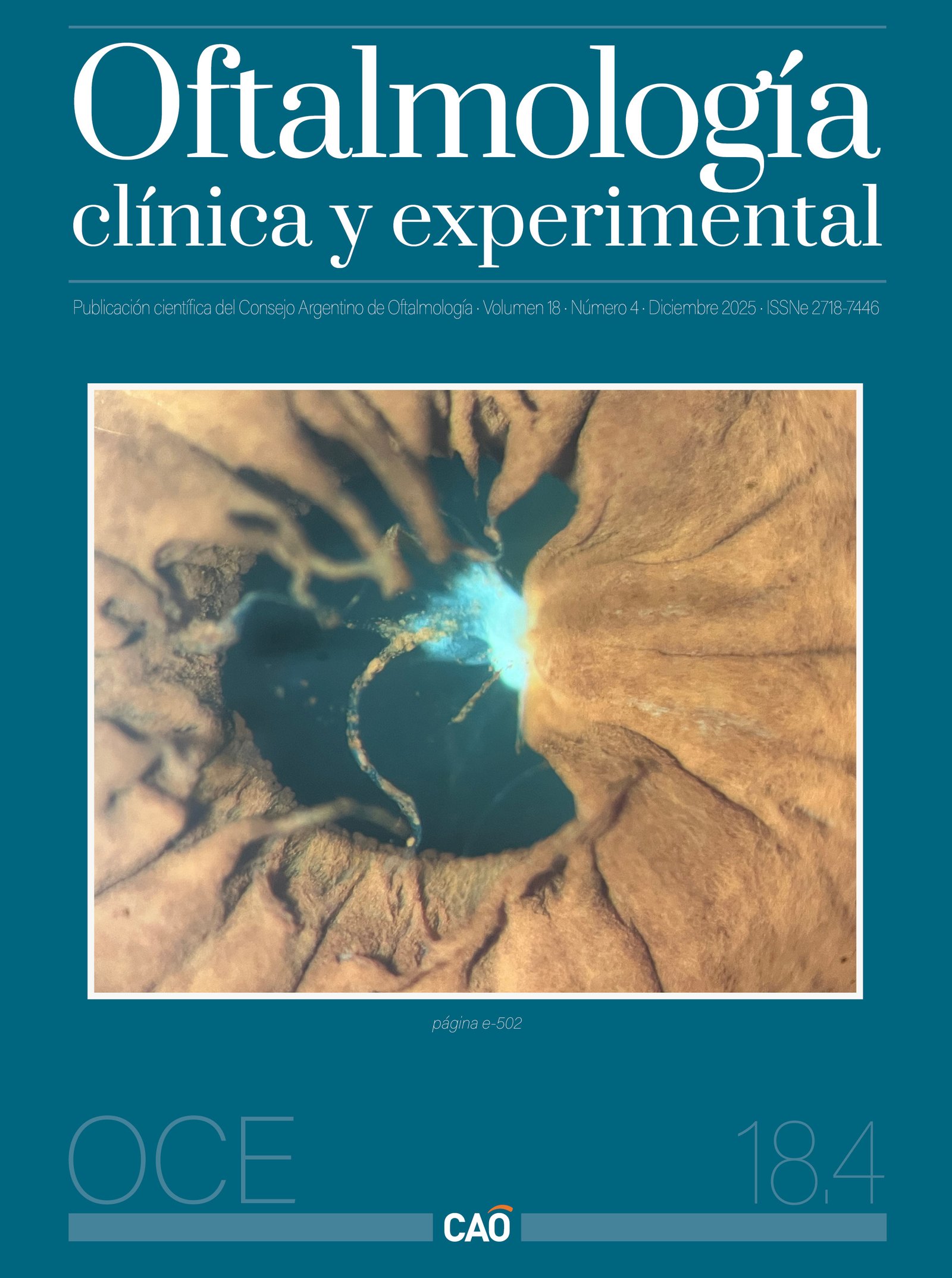
Case report: A 60-year-old man with a family history of ptosis presented with acquired bilateral eyelid ptosis. Visual acuity was 20/20 in both eyes, with a palpebral fissure height of 7 mm in the right eye and 5 mm in the left, and levator function of 15 mm. Bilateral correction was performed by aponeurotic reinsertion of the levator muscle. Due to undercorrection of the right upper eyelid, a reoperation with levator resection was undertaken. Postoperatively, wound dehiscence with granulomatous tissue was observed and managed by margin excision and resuturing. One week later, the patient reported intermittent clear fluid discharge through a pinpoint opening on the upper eyelid, without signs of infection. Detailed examination revealed a fistulous tract between the conjunctiva and the skin, leading to the diagnosis of a postoperative conjunctivo-palpebral fistula. Topical treatment with Cicaplast Baume B5TM (pantenol 5%, madecassoside, tribioma) for 20 days resulted in partial improvement. Subsequently, low-power fractional CO₂ laser treatment was performed around the fistulous site under local anesthesia, achieving complete closure of the tract within 20 days, without recurrence or complications.
Conclusion: Although conjunctivo-palpebral fistula is an uncommon and late complication after ptosis surgery, early recognition and appropriate clinical-surgical management are essential to prevent functional or aesthetic sequelae, as demonstrated in this case.
Downloads
References
1. McInnes CW, Lee-Wing M. Eyelid ptosis. CMAJ 2015; 187(14): 1074. doi: 10.1503/cmaj.140579.
2. Patel K, Carballo S, Thompson L. Ptosis. Dis Mon 2017; 63(3): 74-79. doi: 10.1016/j.disamonth.2016.10.004.
3. Hou D, Tian B, Wang X, Wang Q, Zhu Y. Cause analysis and surgical treatment of aponeurotic ptosis with upper eyelid depression. J Craniofac Surg 2024; 35(7): 1947-1951. doi: 10.1097/SCS.0000000000010155.
4. Mehta S, Belliveau MJ, Oestreicher JH. Oculoplastic surgery. Clin Plast Surg 2013; 40(4): 631-651. doi: 10.1016/j.cps.2013.08.005.
5. Rodrigues C, Carvalho F, Marques M. Upper eyelid blepharoplasty: surgical techniques and results-systematic review and meta-analysis. Aesthetic Plast Surg 2023; 47(5): 1870-1883. doi: 10.1007/s00266-023-03436-6.
6. Kopecký A, Rokohl AC, Heindl LM. The role of the lateral tarsal strip procedure in modern ophthalmic plastic surgery: a review. Front Ophthalmol (Lausanne) 2022; 2: 871964. doi: 10.3389/fopht.2022.871964.
7. Zhang W, Huang Q, Li J. Case report of conjunctival sac fistula after cosmetic lateral canthoplasty. BMC Ophthalmol 2020; 20(1): 127. doi: 10.1186/s12886-020-01402-3.
8. Hou D, Tian B, Zhu Y. Conjunctival fistula after cosmetic lateral canthoplasty. J Craniofac Surg 2022; 33(8): 2578-2580. doi: 10.1097/SCS.0000000000008827.
Downloads
Published
Issue
Section
License
Copyright (c) 2025 Consejo Argentino de Oftalmología

This work is licensed under a Creative Commons Attribution-NonCommercial-NoDerivatives 4.0 International License.
Con esta licencia no se permite un uso comercial de la obra original, ni la generación de obras derivadas. Las licencias Creative Commons permiten a los autores compartir y liberar sus obras en forma legal y segura.
How to Cite


