
Retromode in ophthalmology
current diagnostic applications and clinical perspectives
DOI:
https://doi.org/10.70313/2718.7446.v18.n3.438Keywords:
retinal images, SLO, retromode technique, age related macular degeneration, diabetes retinopathyAbstract
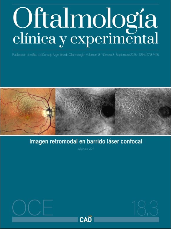
Scanning laser ophthalmoscopes (SLO) were developed with the aim of generating high-contrast images of the chorioretinal layers. Image acquisition through a pinhole aperture enables operation in direct confocal mode, which is employed in the majority of currently available commercial devices.
The ability to laterally displace the pinhole aperture—either to the right or left— as well as the use of an annular aperture, allows for operation in indirect confocal mode.
This indirect confocal mode, termed “RetroMode” by the manufacturer, has been available since 2010 and is exclusively distributed by Nidek in the SLO Nidek F-10 and Mirante SLO devices.
The retromode imaging technique (RMI) provides a perception of depth, enhancing the topographic features of retinal or subretinal pathological structures.
This updated review describes the operating principles of this novel imaging modality and illustrates some of its applications in daily clinical practice.
Downloads
References
1. Mainster MA, Desmettre T, Querques G, Turner PL, Ledesma-Gil G. Scanning laser ophthalmoscopy retroillumination: applications and illusions. Int J Retina Vitreous 2022; 8: 71. doi: 10.1186/s40942-022-00421-0.
2. Cozzi M, Monteduro D, Parrulli S, Corvi F, Zicarelli F, Corradetti G, Sadda SR, Staurenghi G. Sensitivity and specificity of multimodal imaging in characterizing drusen. Ophthalmol Retina 2020; 4(10): 987-995. doi: 10.1016/j.oret.2020.04.013.
3. Yamamoto M, Mizukami S, Tsujikawa A, Miyoshi N, Yoshimura N. Visualization of cystoid macular oedema using a scanning laser ophthalmoscope in the retro-mode. Clin Exp Ophthalmol 2010; 38(1): 27-36. doi: 10.1111/j.1442-9071.2010.02193.x.
4. Ohkoshi K, Tsuiki E, Kitaoka T, Yamaguchi T. Visualization of subthreshold micropulse diode laser photocoagulation by scanning laser ophthalmoscopy in the retro mode. Am J Ophthalmol 2010; 150(6): 856-862. doi: 10.1016/j.ajo.2010.06.022.
5. Savastano MC, Fossataro C, Sadun R, Scupola A, Sammarco MG, Rizzo C, Pafundi PC, Rizzo S. Central serous chorioretinopathy by autofluorescence, enface and SLO-retromode imaging. Life (Basel) 2023; 13(6): 1407. doi: 10.3390/life13061407.
6. Shin YU, Lee BR. Retro-mode imaging for retinal pigment epithelium alterations in central serous chorioretinopathy. Am J Ophthalmol 2012; 154(1): 155-163.e4. doi: 10.1016/j.ajo.2012.01.023.
7. Corradetti G, Byon I, Corvi F, Cozzi M, Staurenghi G, Sadda SR. Retro mode illumination for detecting and quantifying the area of geographic atrophy in non-neovascular age-related macular degeneration. Eye (Lond) 2022; 36(8): 1560-1566. doi: 10.1038/s41433-021-01670-3.
8. Ranetti AE, Stanca HT, Tăbăcaru B, Teodoru A, Munteanu M, Stanca S. Retromode imaging in age-related macular degeneration. Medicina (Kaunas) 2023; 59(4): 647. doi: 10.3390/medicina59040647.
9. Yan Y, Ludwig CA, Liao YJ. Multimodal imaging features of optic disc drusen. Am J Ophthalmol 2021; 225: 18-26. doi: 10.1016/j.ajo.2020.12.023.
10. Ricciotti G, Miere A, Colantuono D, Souied EH. Optic disc drusen using retromode scanning laser ophthalmoscopy. J Neuroophthalmol 2024; 44(3): e479-e480. doi: 10.1097/WNO.0000000000002035.
11. Malmqvist L, Bursztyn L, Costello F, Digre K, Fraser JA, Fraser C, Katz B, Lawlor M, Petzold A, Sibony P, Warner J, Wegener M, Wong S, Hamann S. The optic disc drusen studies consortium recommendations for diagnosis of optic disc drusen using optical coherence tomography. J Neuroophthalmol 2018; 38(3): 299-307. doi: 10.1097/WNO.0000000000000585.
12. Lopez JM, Rabinovich M, Mehanna CJ, Ricciotti G, Crincoli E, Semoun O, Miere A, Souied EH. Retro-mode imaging for the diagnosis of optic disc drusen: a case series. Arch Soc Esp Oftalmol (Engl Ed) 2024; 99(5): 187-194. doi: 10.1016/j.oftale.2024.02.001
Downloads
Published
Issue
Section
License
Copyright (c) 2025 Consejo Argentino de Oftalmología

This work is licensed under a Creative Commons Attribution-NonCommercial-NoDerivatives 4.0 International License.
Con esta licencia no se permite un uso comercial de la obra original, ni la generación de obras derivadas. Las licencias Creative Commons permiten a los autores compartir y liberar sus obras en forma legal y segura.
How to Cite


