CLINICAL CASES
Andrés Germán Alza
Received: August 18th, 2025.
Approved: October 27th, 2025.
Contact
Dr. Andrés Germán Alza
Clínica Privada de Ojos Dr. Enrique Alza
Calle 12, nro. 662
(1900) La Plata, provincia de Buenos Aires
Argentina
+ 54 9 221 4763377
andresalza@hotmail.com
Oftalmol Clin Exp (ISSNe 1851-2658)
2025; 18(4): e499-e509.
DOI: https://doi.org/10.70313/2718.7446.v18.n4.458
Abstract
Objective: To describe a case of persistent pupillary membrane associated with anterior pyramidal cataract and severe ametropia, successfully treated with LASIK refractive surgery, and to analyze the available evidence on the relationship between the two conditions.
Case report: A 55-year-old woman, an emergency physician, with persistent pupillary membrane characterized on biomicroscopy by fine avascular and vascularized strands inserted into a lens with an anterior pyramidal cataract. She had significant myopia and astigmatism, with an initial best-corrected visual acuity of LogMAR 0.10. Preoperative studies were normal. LASIK refractive surgery with an excimer laser (WaveLight® EX500, Alcon) was indicated, with satisfactory results: uncorrected visual acuity of LogMAR 0.10 at one year follow-up, without complications and with full patient satisfaction.
Conclusion: Persistent iridocristalline pupillary membrane is a rare congenital anomaly that rarely contraindicates refractive surgery. There are no documented reports linking it to LASIK, nor is it considered a specific risk factor. In asymptomatic adults with significant ametropia, LASIK may be a valid alternative provided that the usual safety parameters are met.
Keywords: persistent pupillary membrane, congenital anomalies of the iris, anterior pyramidal cataract, refractive surgery, LASIK, myopia, astigmatism.
Membrana pupilar persistente iridocristaliniana monocular asociada a catarata piramidal anterior y ametropía severa: cirugía refractiva con láser de excímero (LASIK)
Resumen
Objetivo: Describir un caso de membrana pupilar persistente asociada a catarata piramidal anterior y ametropía severa, tratada exitosamente con cirugía refractiva LASIK y analizar la evidencia disponible sobre la relación entre ambas entidades.
Caso clínico: Mujer de 55 años, médica de emergencias, con membrana pupilar persistente caracterizada en la biomicroscopía por hebras finas avasculares y vascularizadas insertadas en un cristalino con una catarata piramidal anterior. Presentaba miopía y astigmatismo —ambos significativos— con agudeza visual mejor corregida inicial de LogMAR 0,10. Los estudios prequirúrgicos fueron normales. Se indicó cirugía refractiva LASIK con excímer láser (WaveLight® EX500, Alcon), obteniéndose resultados satisfactorios: agudeza visual no corregida de LogMAR 0,10 en un año de seguimiento, sin complicaciones y con plena satisfacción de la paciente.
Conclusión: La membrana pupilar persistente iridocristaliniana es una anomalía congénita poco frecuente que raramente contraindica la cirugía refractiva. No se han documentado reportes que la relacionen con LASIK, ni se considera un factor de riesgo específico. En adultos asintomáticos con ametropías significativas, el LASIK puede constituir una alternativa válida siempre que se cumplan los parámetros de seguridad habituales.
Palabras clave: membrana pupilar persistente, anomalías congénitas del iris, catarata piramidal anterior, cirugía refractiva, LASIK, miopía, astigmatismo.
Membrana pupilar iridocristaliniana monocular persistente associada a catarata piramidal anterior e ametropia grave: cirurgia refrativa com laser excimer (LASIK)
Resumo
Objetivo: Descrever um caso de membrana pupilar iridocristaliniana persistente associada à catarata piramidal anterior e ametropia grave, tratado com sucesso por meio de cirurgia refrativa LASIK, e analisar as evidências disponíveis sobre a relação entre ambas as condições.
Caso clínico: Mulher, médica de emergência de 55 anos, com membrana pupilar iridocristaliniana persistente caracterizada à biomicroscopia por finos filamentos avasculares e vascularizados inseridos em um cristalino com catarata piramidal anterior. A paciente apresentava miopia e astigmatismo significativos, com acuidade visual inicial corrigida de 0,10 LogMAR. Os exames pré-operatórios foram normais. Foi indicada a cirurgia refrativa LASIK com laser excimer (WaveLight® EX500, Alcon), que apresentou resultados satisfatórios: acuidade visual não corrigida de 0,10 LogMAR após um ano de acompanhamento, sem complicações e com total satisfação da paciente.
Conclusão: A membrana iridocristaliniana persistente é uma anomalia congênita rara que raramente contraindica a cirurgia refrativa. Não há relatos documentados que a associem ao LASIK, nem é considerada um fator de risco específico. Em adultos assintomáticos com erros refrativos significativos, o LASIK pode ser uma alternativa válida, desde que os parâmetros de segurança padrão sejam atendidos.
Palavras-chave: membrana pupilar iridocristaliniana persistente, anomalias congênitas da íris, catarata piramidal anterior, cirurgia refrativa, LASIK, miopía, astigmatismo.
Introduction
Persistent pupillary membrane (PPM) is an embryological remnant of the pupillary vascular membrane, which normally regresses before birth. On slit-lamp examination, it appears as fine strands extending from the iris collarette towards the pupillary centre or, as in this case, to the anterior lens capsule, giving rise to an anterior pyramidal cataract.
It is usually asymptomatic and diagnosed incidentally during routine examinations, although it may be associated with other anterior segment anomalies or compromise the visual axis. The objective of this work is to present a case of PPM with severe unilateral refractive error, successfully treated with LASIK refractive surgery.
Case report
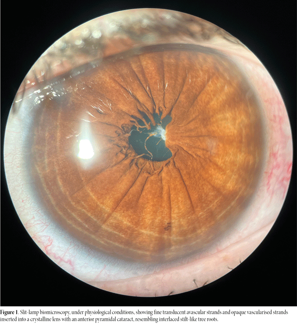
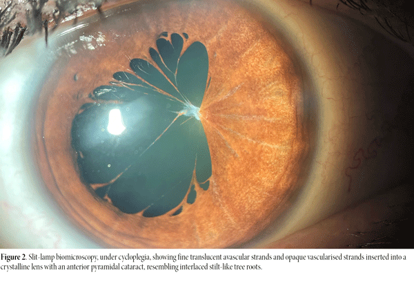
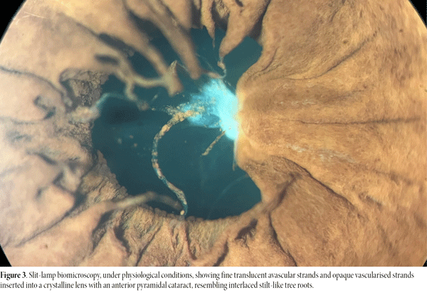
A 55-year-old female emergency physician was diagnosed with persistent pupillary membrane (PPM). Slit-lamp biomicroscopy under cycloplegia revealed fine translucent avascular strands and opaque vascularised strands inserted into the crystalline lens with an anterior pyramidal cataract, resembling interlaced stilt-like roots (Figs. 1-3).
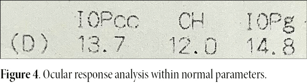
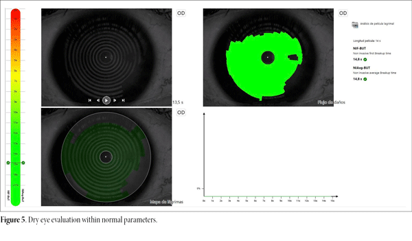
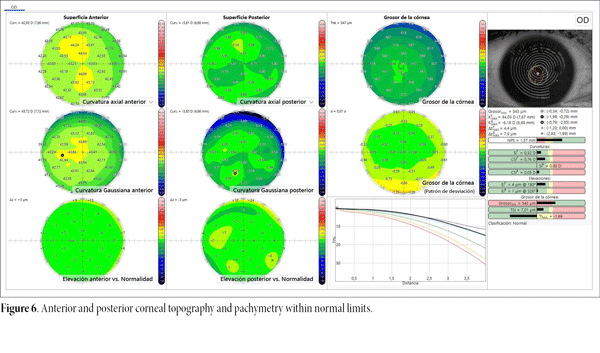
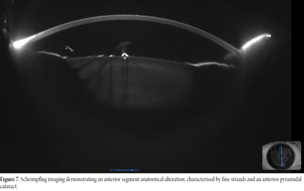
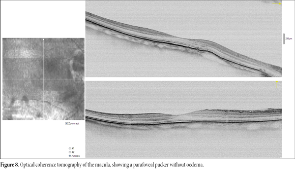
Preoperatively, her uncorrected visual acuity was LogMAR 1.33. Refraction testing revealed a best-corrected visual acuity of LogMAR 0.10, with a refractive error of -5.75 D sphere and -1.00 D cylinder at 12°. Preoperative assessments —including ocular response analysis (Fig. 4), dry eye evaluation (Fig. 5), corneal topography and pachymetry (Fig. 6)— were within normal limits. Scheimpflug imaging demonstrated an anterior segment anatomical alteration characterised by the strands and the pyramidal cataract (Fig. 7). Optical coherence tomography revealed a normal optic disc, while macular imaging showed a parafoveal pucker without oedema (Fig. 8).
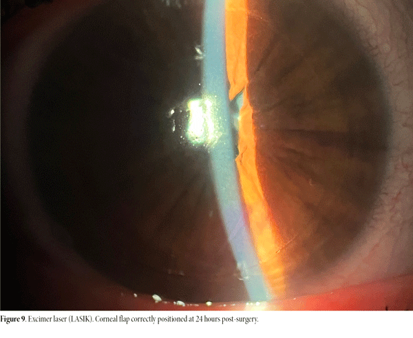
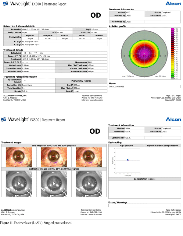
Excimer laser refractive surgery (LASIK, WaveLight® EX500, Alcon) was indicated (Figs. 9-11). The procedure was successful, achieving an uncorrected visual acuity of LogMAR 0.10 at one-year follow-up, with no complications and full patient satisfaction.
 ç
ç
Discussion
PPM is a congenital anomaly secondary to incomplete regression of the tunica vasculosa, the fetal vascular network that nourishes the developing crystalline lens. Although most cases are sporadic and asymptomatic, an autosomal dominant inheritance pattern has been described in patients with associated anterior segment anomalies1.
Clinically, it presents as filaments extending from the iris collarette towards the pupil, cornea, or anterior capsule. The differential diagnosis includes pupillary abnormalities such as coloboma, pseudoacorea in Axenfeld-Rieger syndrome2-3, trauma4, accessory iris membrane5, and post-inflammatory synechiae.
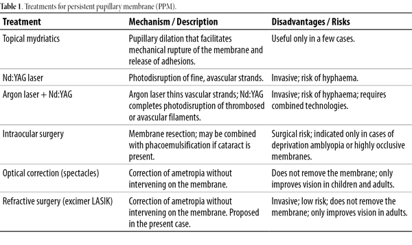
Management depends on age and the degree of visual compromise (Table 1). Options include optical correction with spectacles6, topical atropine7, surgical removal of the membrane in cases of visual axis occlusion7-8, or phacoemulsification when associated with cataract7. Nd:YAG laser therapy has also been reported9, either alone or combined with argon laser, the latter being useful for thinning the membrane prior to Nd:YAG photodisruption10.
Regarding LASIK, no previous reports have been found linking it to PPM. The refractive surgery literature focuses on pupil size and its influence on postoperative visual quality, without identifying PPM as a contraindication or risk factor. In practice, it rarely alters the indication or outcomes, except in cases of dense membranes that obstruct the visual axis.
In the present case, the severe unilateral ametropia compromised the patient’s daily visual performance, justifying the indication for LASIK, which yielded favourable results without complications.
Conclusion
PPM is an uncommon congenital anomaly that rarely compromises the indication for refractive surgery. The association between PPM and LASIK has not been reported in the literature, as it represents a rare condition, usually without functional relevance or impact on refractive surgery. In adults with significant ametropia and no visual axis obstruction, LASIK may constitute a valid and safe alternative, provided that standard surgical safety parameters are observed.
References
Figures
Figure 1. Slit-lamp biomicroscopy, under physiological conditions, showing fine translucent avascular strands and opaque vascularised strands inserted into a crystalline lens with an anterior pyramidal cataract, resembling interlaced stilt-like tree roots.
Figure 2. Slit-lamp biomicroscopy, cycloplegia, showing fine translucent avascular strands and opaque vascularised strands inserted into a crystalline lens with an anterior pyramidal cataract, resembling interlaced stilt-like tree roots.
Figure 3. Slit-lamp biomicroscopy, under physiological conditions, showing fine translucent avascular strands and opaque vascularised strands inserted into a crystalline lens with an anterior pyramidal cataract, resembling interlaced stilt-like tree roots.
Figure 4. Ocular response analysis within normal parameters.
Figure 5. Dry eye evaluation within normal parameters.
Figure 6. Anterior and posterior corneal topography and pachymetry within normal limits.
Figure 7. Scheimpflug imaging demonstrating an anterior segment anatomical alteration, characterised by fine strands and an anterior pyramidal cataract.
Figure 8. Optical coherence tomography of the macula, showing a parafoveal pucker without oedema.
Figure 9. Excimer laser (LASIK). Corneal flap correctly positioned at 24 hours post-surgery.
Figure 10A. Excimer laser (LASIK). Corneal surface marking and pachymetry.
Figure 10B. Excimer laser (LASIK). Corneal flap creation.
Figura 10C. Excimer laser (LASIK). Excimer laser apllication, pachymetry, and repositioning of the corneal flap.
Figure 11. Excimer laser (LASIK). Surgical protocol used.